 |
| Wilhelm Röntgen Public Domain, Link |
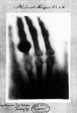
In 1895, Wilhelm Röntgen, a physics professor in Bavaria, took the very first X-ray image of the human body. He called it a “röntgenogram.” It was an image of his wife’s hand.
He didn't know what impact the imaging might have on his wife. No one knew. Röntgen’s X-ray used 1,500 times the dosage of radiation used by modern machines, and it took 90 minutes of exposure to get the full effect.
Maybe Mrs. Röntgen might have sensed something because when she saw the skeletal image of her hand, she exclaimed, “I have seen my death!” It took many years before people made the connection between frequent exposure to X-rays and higher rates of cancer.
The invention was a breakthrough as it allowed views of the human body without surgery. In less than a year, the Glasgow Royal Infirmary became the first hospital to set up a radiology department. Doctors and experimenting scientists X-rayed all kinds of things found in bodies: a penny in a child’s throat, kidney stones, bone fractures, and shrapnel in a soldier.
 |
| Natural color x-ray photogram by NickSpiker CC BY-SA 4.0, Link |
No comments:
Post a Comment